812 results filtered with: Pictures

- Pictures
- Online
Dissections of diseased lungs affected by pneumonia and tuberculosis: two figures. Chromolithograph by W. Gummelt, ca. 1897.
Gummelt, W.Date: [1897?]Reference: 577571i
- Pictures
- Online
Dissections of the stomach and rumination system of a cow: two figures. Chromolithograph by H.J. Ruprecht, 1877.
Ruprecht, H. J.Date: [1877]Reference: 578973i
- Pictures
- Online
Dissections of the upper jaw and the sinuses (?): three figures. Colour lithograph by G.H. Ford, 1865.
Ford, G. H. (George Henry)Date: 1865Reference: 580342i
- Pictures
- Online
Dissections of diseased livers: two figures showing symptoms of cholangitis and cholecystitis. Chromolithograph by W. Gummelt, ca. 1897.
Gummelt, W.Date: [1897?]Reference: 577056i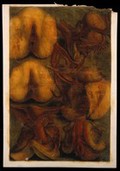
- Pictures
- Online
Dissections of the female urogenital system: six figures. Colour mezzotint by J. F. Gautier d'Agoty after himself, 1754.
Gautier Dagoty, 1717-1785.Date: [1754]Reference: 572095i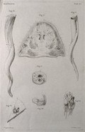
- Pictures
- Online
Dissections of an elephant's trunk and tusks: seven figures. Etching by R. Vinkeles 1787/1800 (?), after P. Camper, 1774.
Camper, Petrus, 1722-1789.Date: [1787/1800?]Reference: 571093i
- Pictures
- Online
Dissections of an elephant's foot and tail: seven figures. Etching by R. Vinkeles 1787/1800 (?), after P. Camper, 1774.
Camper, Petrus, 1722-1789.Date: [1787/1800?]Reference: 571094i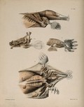
- Pictures
- Online
Dissections of a mole: four figures, showing the muscles of the head and limbs. Lithograph by F.W. Brookman, 1880/1900?.
Brookman, F. W.Date: [1880/1900?]Reference: 571450i
- Pictures
- Online
Dissections of a diseased layrnx and pharynx and part of the duodenum: two figures. Chromolithograph by W. Gummelt, ca. 1897.
Gummelt, W.Date: [1897?]Reference: 577249i
- Pictures
- Online
Dissections of diseased intestines and mesenteries, affected by necrosis, ulcers and tuberculosis: three figures. Chromolithograph by W. Gummelt, ca. 1897.
Gummelt, W.Date: [1897?]Reference: 577380i
- Pictures
- Online
Dissections of chicks of various species: eleven figures, including details of the feet. Lithograph by J. Erxleben (?), 1840/1860?.
Erxleben, James, active 1839-1852.Date: [1840/1860?]Reference: 571033i
- Pictures
- Online
Dissections of diseased livers: two figures showing symptoms of amyloidosis hepatis and haemo-siderosis hepatis. Chromolithograph by W. Gummelt, ca. 1897.
Gummelt, W.Date: [1897?]Reference: 577059i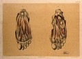
- Pictures
- Online
Dissections of the underside of the foot, showing the muscles and blood vessels: two figures. Colour lithograph by G.H. Ford, 1867.
Ford, G. H. (George Henry)Date: 1867Reference: 580546i
- Pictures
- Online
Dissections of diseased livers: four figures showing symptoms caused by hepatitis, syphilis, anfioma cavernosum and pylephlebitis. Chromolithograph by W. Gummelt, ca. 1897.
Gummelt, W.Date: [1897?]Reference: 577054i
- Pictures
- Online
Dissections of femoral arteries affected by calcification, with illustrations of subperitoneal phlebectasia and rectal rectal hemorrhoids. Chromolithograph by W. Gummelt, ca. 1897.
Gummelt, W.Date: [1897?]Reference: 577242i
- Pictures
- Online
Dissections of diseased hearts: three figures, including hearts affected by forms of endocarditis and myocardial haemorrhage. Chromolithograph by W. Gummelt, ca. 1897.
Gummelt, W.Date: [1897?]Reference: 577233i
- Pictures
- Online
Dissections of a mole: sixteen figures, showing the bones and musculature of the animal. Lithograph by E. Wilson and F.W. Brookman, 1880/1900?.
Brookman, F. W.Date: [1880/1900?]Reference: 571432i- Pictures
Dissections of diseased lungs and thyroid gland, affected by diseases including tuberculosis and cancer (?): four figures. Chromolithograph by W. Gummelt, ca. 1897.
Gummelt, W.Date: [1897?]Reference: 577585i
- Pictures
- Online
Dissections of a mole: thirteen figures, including the musculature of the head, neck, legs and feet. Lithograph by R. Mintern after F.W. Brookman, 1880/1900?.
Brookman, F. W.Date: [1880/1900?]Reference: 571437i
- Pictures
- Online
Dissections of the arm and hand; four figures, with the arteries and blood vessels indicated in red. Coloured lithograph by J. Roux, 1822.
Roux, Jakob Wilhelm Christian, 1775-1831.Date: [1822]Reference: 579759iPart of: Tiedemann, Friedrich, 1781-1861.
- Pictures
- Online
Dissections of the head of a mole: three figures, showing the musculature of the animal's head and neck. Lithograph by E. Wilson and F.W. Brookman, 1880/1900?.
Brookman, F. W.Date: [1880/1900?]Reference: 571433i
- Pictures
- Online
Dissections of an ulcerated stomach caused by syphilis, and a severe case of ulcerated proctitis of the rectum (?): two figures. Chromolithograph by W. Gummelt, ca. 1897.
Gummelt, W.Date: [1897?]Reference: 577375i
- Pictures
- Online
Dissections of the male anus and urogenital system: two figures, with the arteries and blood vessels indicated in red. Coloured lithograph by J. Roux, 1822.
Roux, Jacob Chr.Date: [1822]Reference: 57976iPart of: Tiedemann, Friedrich, 1781-1861.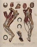
- Pictures
- Online
Dissections of a horse's leg and foot: eleven figures, showing the muscles and blood-vessels of the foot, with examples of horseshoes. Coloured engraving by J. Pass after Harguinier, 1805.
Harguinier, active 1763-1768.Date: 1805Reference: 570700i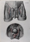
- Pictures
- Online
Dissections of the male genitals, upper thighs and pelvic region: two figures, with the arteries, blood vessels and veins indicated in red and blue. Coloured lithograph by J. Roux, 1822.
Roux, Jacob Chr.Date: [1822]Reference: 579771iPart of: Tiedemann, Friedrich, 1781-1861.